1. Ενδείξεις
1). Τα σοβαρά συντριπτικά κατάγματα έχουν εμφανή μετατόπιση και η αρθρική επιφάνεια της περιφερικής κερκίδας καταστρέφεται.
2). Η χειροκίνητη ανάταξη απέτυχε ή η εξωτερική οστεοσύνθεση απέτυχε να διατηρήσει τη ανάταξη.
3).Παλαιά κατάγματα.
4). Κάταγμα πλημμελούς ή μη πώρωσης. οστό που υπάρχει στην ημεδαπή και στο εξωτερικό
2. Αντενδείξεις
Ηλικιωμένοι ασθενείς που δεν είναι κατάλληλοι για χειρουργική επέμβαση.
3. Τεχνική εξωτερικής στερέωσης
1. Εξωτερική οστεοσύνθεση διααρθρικής άρθρωσης για την αποκατάσταση καταγμάτων περιφερικής κερκίδας
Θέση και προεγχειρητική προετοιμασία:
·Αναισθησία βραχιόνιου πλέγματος
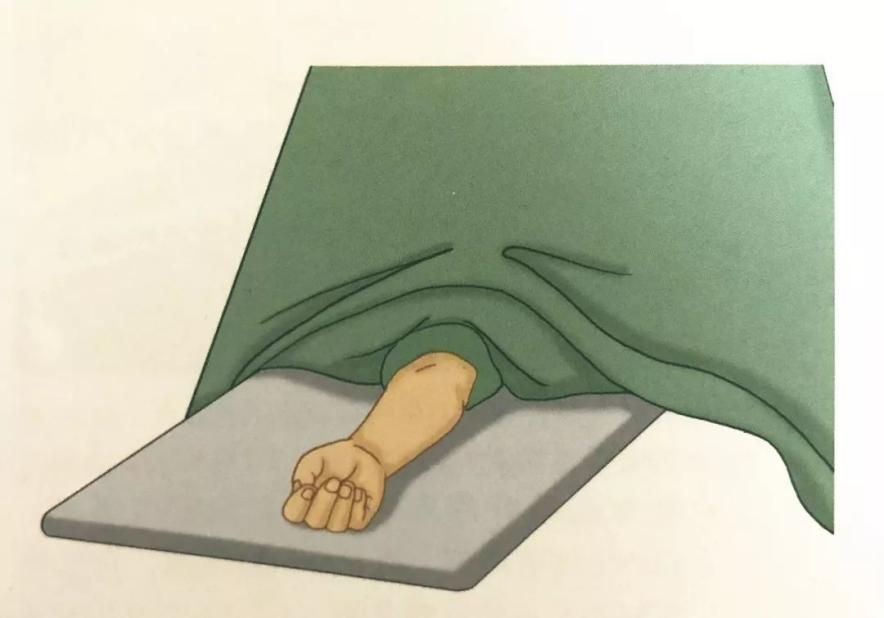
·Υπτία θέση με το πάσχον άκρο σε επίπεδη θέση πάνω στο διαφανές άγκιστρο δίπλα στο κρεβάτι
·Εφαρμόστε ένα αιμοστατικό επίδεσμο στο 1/3 του άνω βραχίονα
·Παρατήρηση προοπτικής
Χειρουργική τεχνική
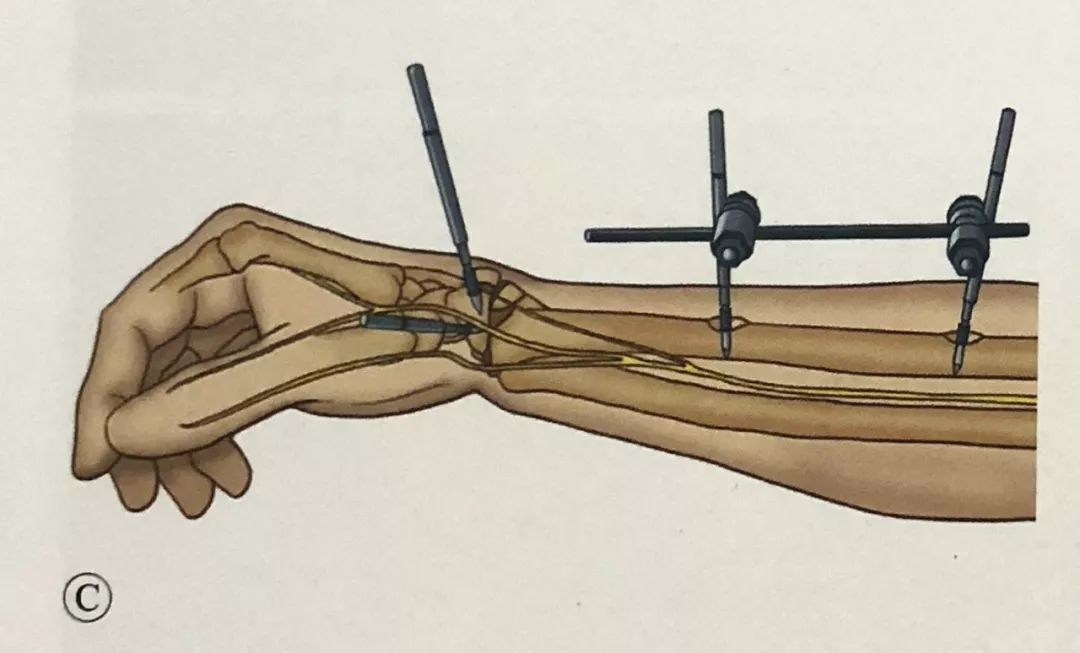
Εισαγωγή μετακαρπικής βίδας:
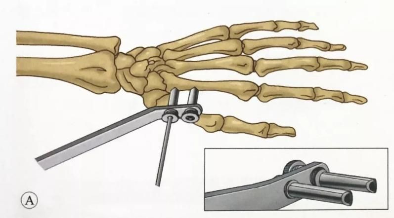
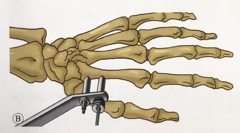
Η πρώτη βίδα βρίσκεται στη βάση του δεύτερου μετακαρπίου οστού. Γίνεται δερματική τομή μεταξύ του εκτείνοντος τένοντα του δείκτη και του ραχιαίου μεσοοστέου μυός του πρώτου οστού. Ο μαλακός ιστός διαχωρίζεται απαλά με χειρουργική λαβίδα. Το χιτώνιο προστατεύει τον μαλακό ιστό και εισάγεται μια βίδα Schanz 3 mm. Βίδες
Η κατεύθυνση της βίδας είναι 45° ως προς το επίπεδο της παλάμης ή μπορεί να είναι παράλληλη με το επίπεδο της παλάμης.
Χρησιμοποιήστε τον οδηγό για να επιλέξετε τη θέση της δεύτερης βίδας. Μια δεύτερη βίδα 3 mm μπήκε στο δεύτερο μετακάρπιο.
Η διάμετρος της καρφίτσας στερέωσης του μετακαρπίου δεν πρέπει να υπερβαίνει τα 3 mm. Η καρφίτσα στερέωσης βρίσκεται στο εγγύς 1/3. Για ασθενείς με οστεοπόρωση, η πιο εγγύς βίδα μπορεί να διαπεράσει τρία στρώματα του φλοιού (το δεύτερο μετακάρπιο οστό και το μισό φλοιό του τρίτου μετακαρπίου οστού). Με αυτόν τον τρόπο, η βίδα. Ο μακρύς βραχίονας στερέωσης και η μεγάλη ροπή στερέωσης αυξάνουν τη σταθερότητα της καρφίτσας στερέωσης.
Τοποθέτηση ακτινικών βιδών:
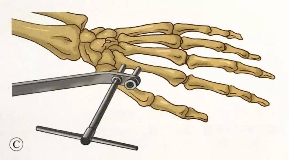
Κάντε μια τομή δέρματος στην πλάγια άκρη της κερκίδας, μεταξύ του βραχιονιακού μυός και του εκτείνοντα μυός του κερκιδικού καρπού, 3 cm πάνω από το εγγύς άκρο της γραμμής του κατάγματος και περίπου 10 cm εγγύς της άρθρωσης του καρπού, και χρησιμοποιήστε αιμοστάτη για να διαχωρίσετε αμβλύς τον υποδόριο ιστό από την επιφάνεια του οστού. Λαμβάνεται μέριμνα για την προστασία των επιφανειακών κλάδων του κερκιδικού νεύρου που πορεύονται σε αυτήν την περιοχή.
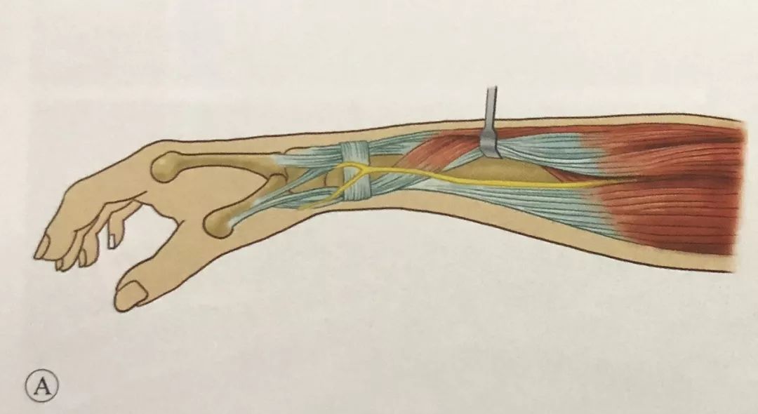
Στο ίδιο επίπεδο με τις μετακαρπικές βίδες, τοποθετήθηκαν δύο βίδες Schanz 3 mm υπό την καθοδήγηση του οδηγού μαλακών ιστών προστασίας μανικιού.
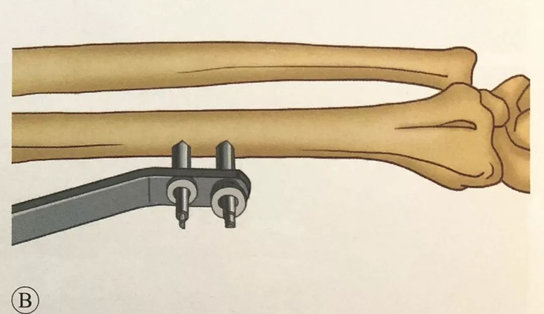
·.Μείωση και στερέωση καταγμάτων:
·. Χειροκίνητη ανάταξη έλξης και ακτινοσκόπηση C-arm για τον έλεγχο της ανάταξης του κατάγματος.
· Η εξωτερική στερέωση κατά μήκος της άρθρωσης του καρπού δυσχεραίνει την πλήρη αποκατάσταση της παλαμιαίας γωνίας κλίσης, επομένως μπορεί να συνδυαστεί με καρφίτσες Kapandji για να βοηθήσει στην ανάταξη και τη στερέωση.
· Για ασθενείς με κατάγματα ακτινωτού στυλοειδούς, μπορεί να χρησιμοποιηθεί στερέωση με σύρμα Kirschner με ακτινωτό στυλοειδές.
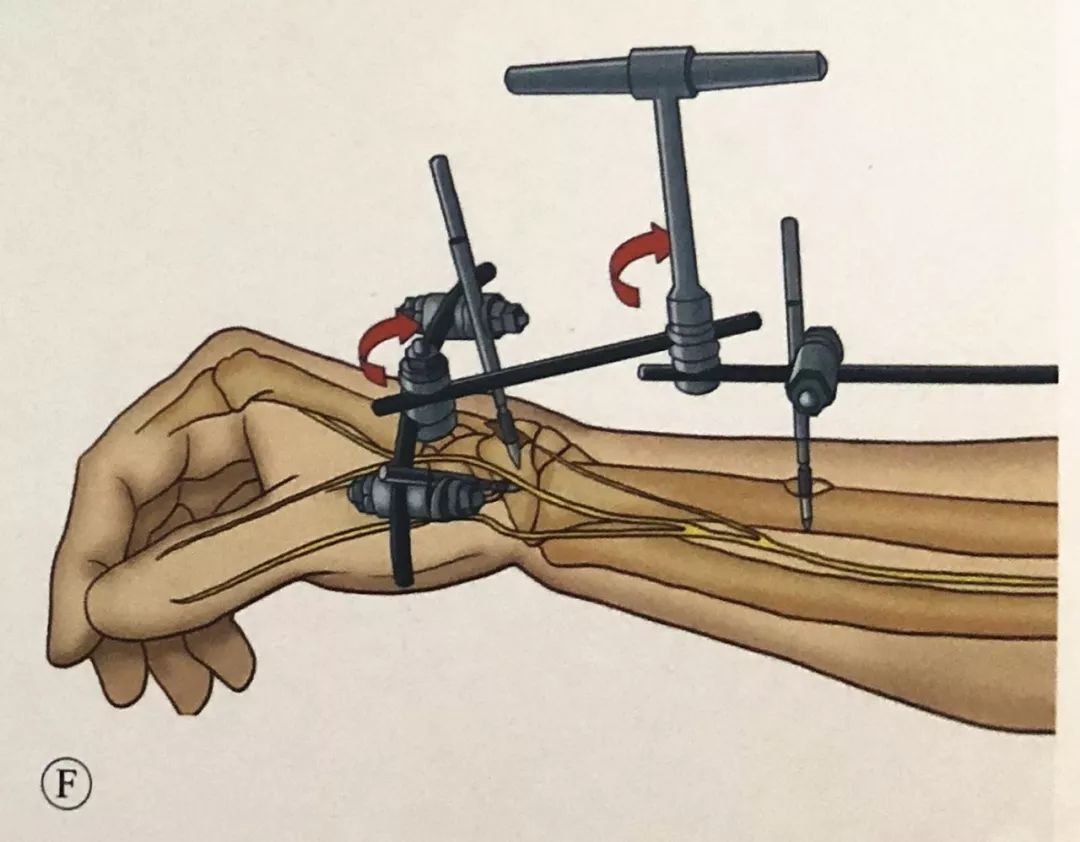
·.Διατηρώντας την ανάταξη, συνδέστε το εξωτερικό οστεοσύνθετο και τοποθετήστε το κέντρο περιστροφής του εξωτερικού οστεοσύνθετου στον ίδιο άξονα με το κέντρο περιστροφής της άρθρωσης του καρπού.
·.Προσοοπίσθια και πλάγια ακτινοσκόπηση, έλεγχος εάν το μήκος της κερκίδας, η γωνία κλίσης της παλάμης και η γωνία απόκλισης της ωλένης έχουν αποκατασταθεί και ρύθμιση της γωνίας στερέωσης μέχρι η μείωση του κατάγματος να είναι ικανοποιητική.
· Δώστε προσοχή στην έλξη του εξωτερικού οστεοσταθεροποιητή, η οποία προκαλεί ιατρογενή κατάγματα στις βίδες του μετακαρπίου.
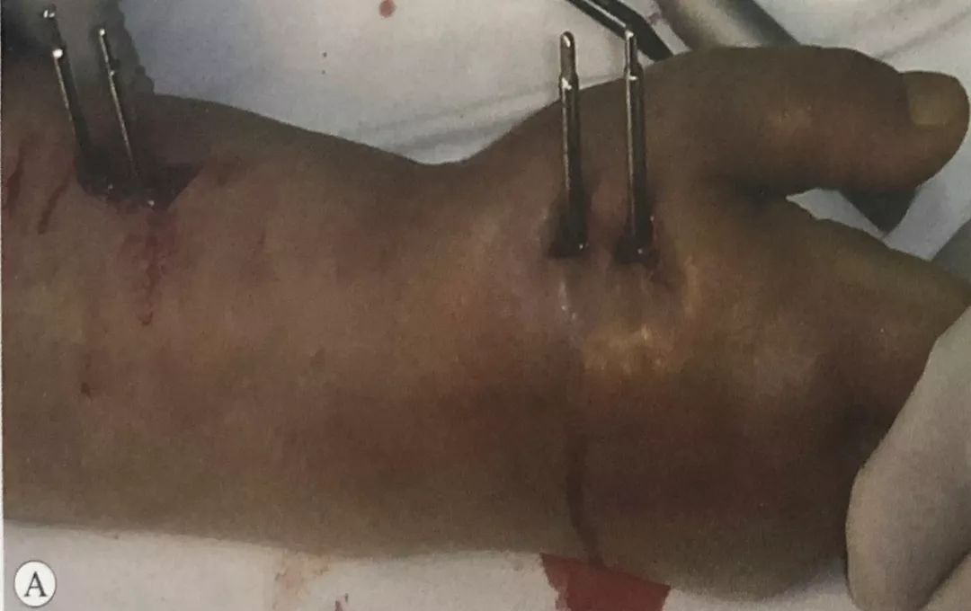
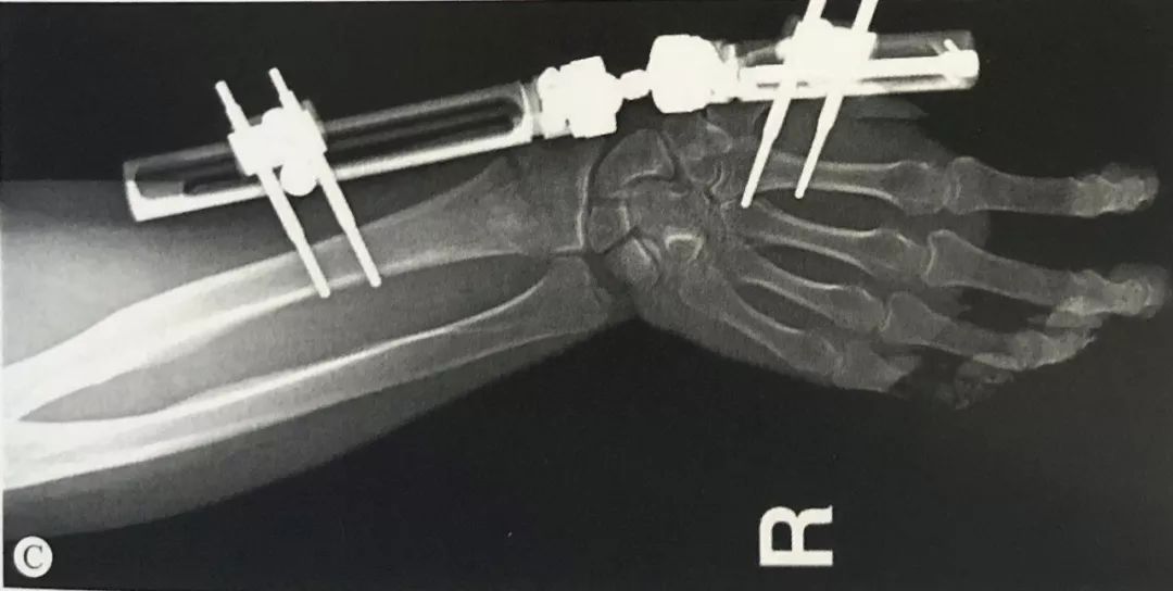
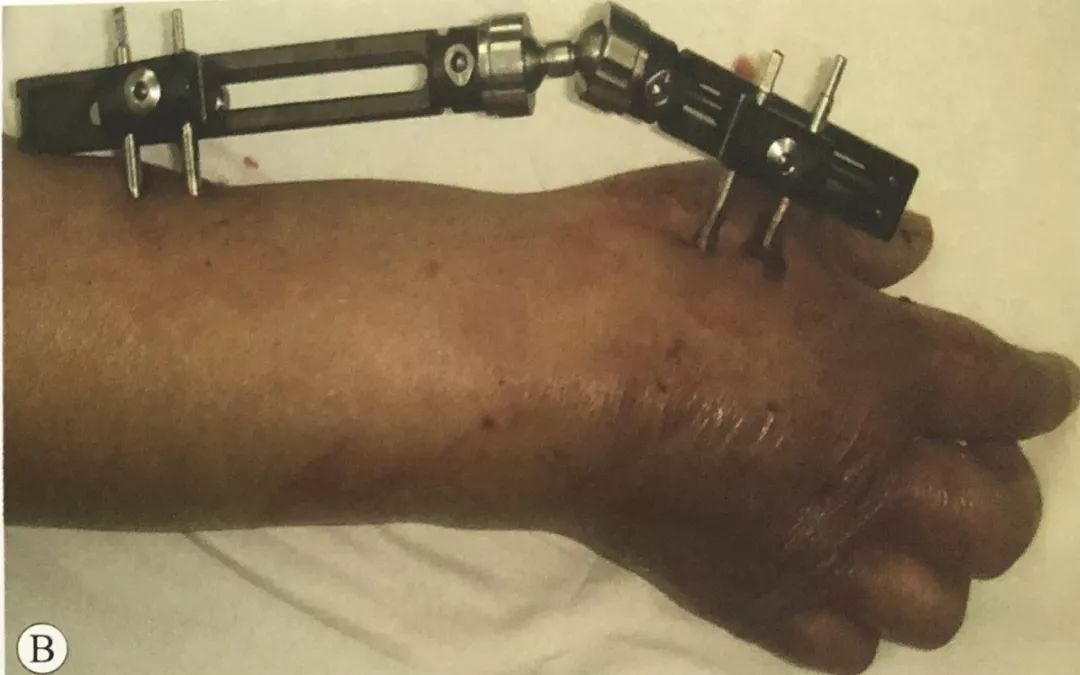
Κάταγμα περιφερικής κερκίδας σε συνδυασμό με διαχωρισμό της περιφερικής κερκιδοωλένιας άρθρωσης (DRUJ):
·.Οι περισσότερες DRUJ μπορούν να αναταχθούν αυθόρμητα μετά από ανάταξη της περιφερικής κερκίδας.
·.Εάν το DRUJ εξακολουθεί να είναι διαχωρισμένο μετά τη μείωση της περιφερικής κερκίδας, χρησιμοποιήστε χειροκίνητη συμπίεση και χρησιμοποιήστε την πλευρική στερέωση με ράβδο του εξωτερικού βραχίονα.
· Ή χρησιμοποιήστε σύρματα Κ για να διαπεράσετε το DRUJ στην ουδέτερη ή ελαφρώς υπτιασμένη θέση.
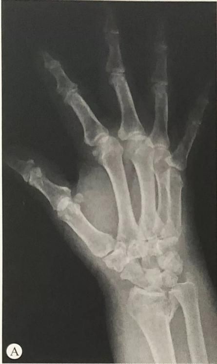
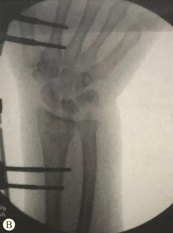
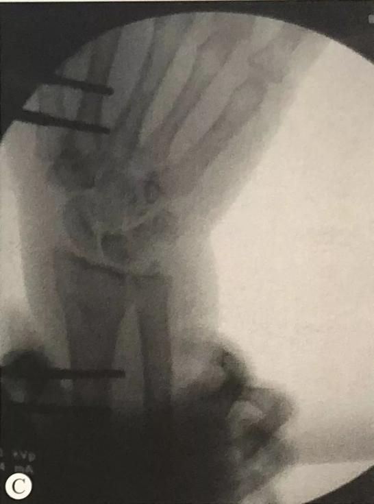
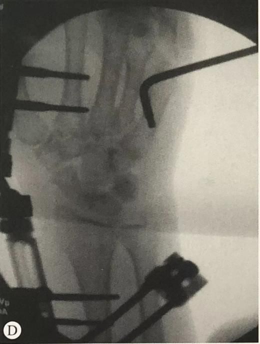
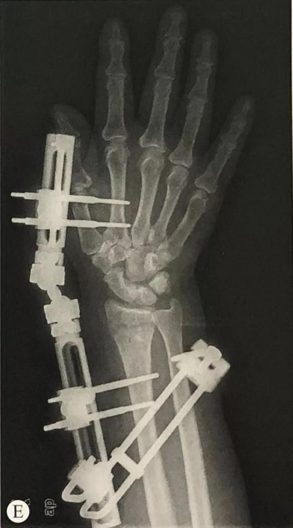
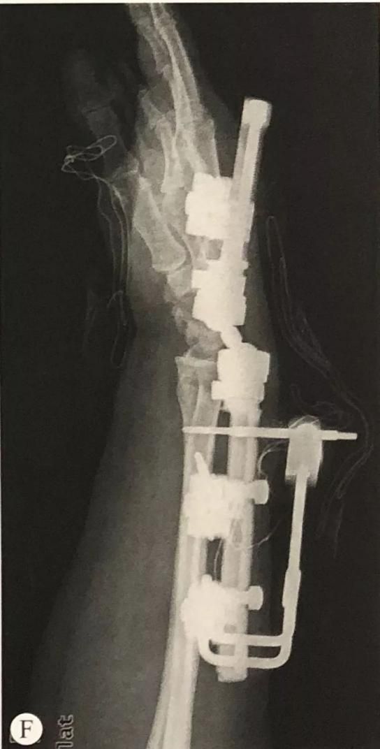
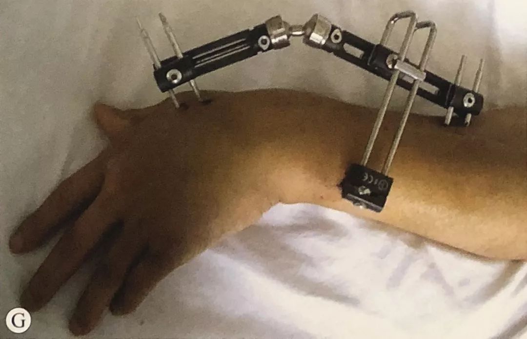
Κάταγμα περιφερικού άκρου της κερκίδας σε συνδυασμό με κάταγμα στυλοειδούς της ωλένης: Ελέγξτε τη σταθερότητα της άρθρωσης του κάτω άκρου (DRUJ) σε πρηνισμό, ουδέτερη θέση και υπτιασμό του αντιβραχίου. Εάν υπάρχει αστάθεια, μπορεί να χρησιμοποιηθεί υποβοηθούμενη οστεοσύνθεση με σύρματα Kirschner, αποκατάσταση του συνδέσμου TFCC ή η αρχή της ζώνης τάσης για την οστεοσύνθεση της στυλοειδούς απόφυσης της ωλένης.
Αποφύγετε το υπερβολικό τράβηγμα:
· Ελέγξτε εάν τα δάχτυλα του ασθενούς μπορούν να εκτελέσουν πλήρεις κινήσεις κάμψης και έκτασης χωρίς εμφανή τάση· συγκρίνετε τον ακτινωτού μήκους αρθρικό χώρο και τον μεσοκαρπιαίο αρθρικό χώρο.
·Ελέγξτε αν το δέρμα στο κανάλι του νυχιού είναι πολύ σφιχτό. Εάν είναι πολύ σφιχτό, κάντε μια κατάλληλη τομή για να αποφύγετε τη μόλυνση.
·Ενθαρρύνετε τους ασθενείς να κινούν τα δάχτυλά τους νωρίς, ειδικά την κάμψη και έκταση των μετακαρποφαλαγγικών αρθρώσεων των δακτύλων, την κάμψη και έκταση του αντίχειρα και την απαγωγή.
2. Οστεοσύνθεση καταγμάτων περιφερικής κερκίδας με εξωτερικό οστεοσύνθεση που δεν διασχίζει την άρθρωση:
Θέση και προεγχειρητική προετοιμασία: Όπως και πριν.
Χειρουργικές τεχνικές:
Οι ασφαλείς περιοχές για την τοποθέτηση του σύρματος K στην ραχιαία πλευρά της περιφερικής κερκίδας είναι: εκατέρωθεν του φύματος Lister, εκατέρωθεν του τένοντα του μακρού εκτείνοντα του αντίχειρα και μεταξύ του τένοντα του κοινού εκτείνοντα των δακτύλων και του τένοντα του μικρού εκτείνοντα των δακτύλων.
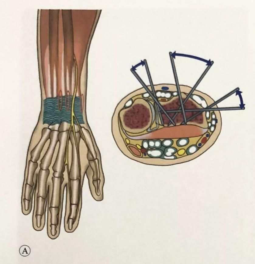
Με τον ίδιο τρόπο, δύο βίδες Schanz τοποθετήθηκαν στον ακτινικό άξονα και συνδέθηκαν με μια μπιέλα.
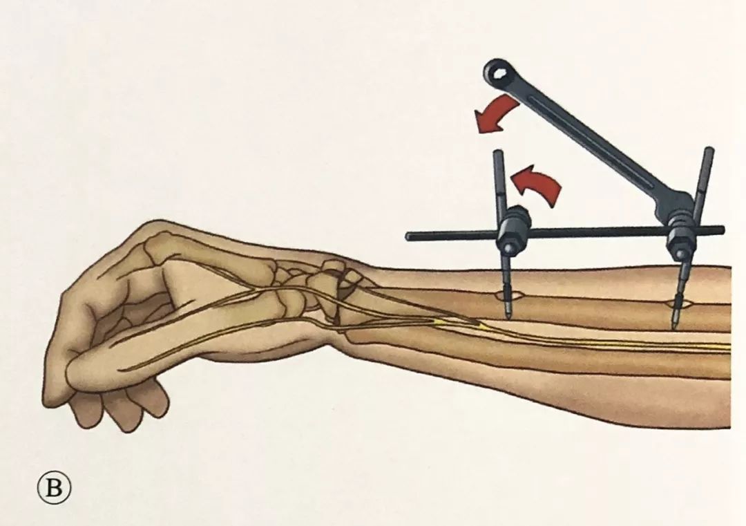
Μέσω της ζώνης ασφαλείας, εισήχθησαν δύο βίδες Schanz στο θραύσμα του περιφερικού κατάγματος της κερκίδας, μία από την κερκιδική πλευρά και μία από την ραχιαία πλευρά, με γωνία 60° έως 90° μεταξύ τους. Η βίδα θα πρέπει να συγκρατεί τον ετερόπλευρο φλοιό και πρέπει να σημειωθεί ότι η άκρη της βίδας που εισάγεται στην κερκιδική πλευρά δεν μπορεί να περάσει από την σιγμοειδή εντομή και να εισέλθει στην περιφερική κερκιδοωλενική άρθρωση.
Συνδέστε τη βίδα Schanz στην κερκίδα με μια καμπύλη σύνδεση.
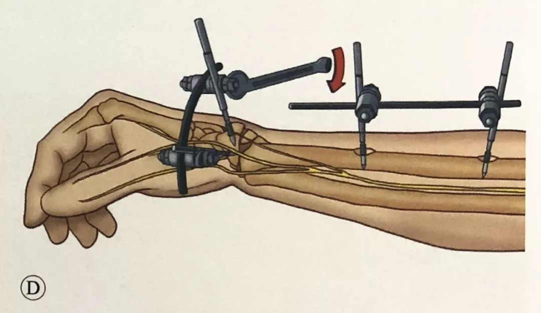
Χρησιμοποιήστε μια ενδιάμεση μπιέλα για να συνδέσετε τα δύο σπασμένα μέρη και προσέξτε να μην κλειδώσετε προσωρινά το τσοκ. Με τη βοήθεια του ενδιάμεσου συνδέσμου, το περιφερικό θραύσμα μειώνεται.
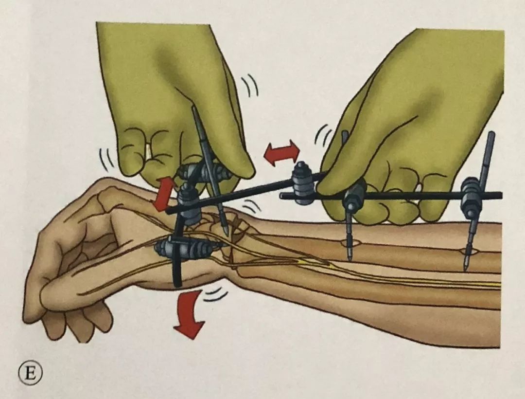
Μετά την επαναφορά, ασφαλίστε το τσοκ στη μπιέλα για να ολοκληρώσετε την τελικήστερέωση.
Η διαφορά μεταξύ εξωτερικής οστεοσύνθεσης χωρίς άνοιγμα αρθρώσεων και εξωτερικής οστεοσύνθεσης διασταυρούμενων αρθρώσεων:
Επειδή μπορούν να τοποθετηθούν πολλαπλές βίδες Schanz για την ολοκλήρωση της ανάταξης και της στερέωσης οστικών θραυσμάτων, οι χειρουργικές ενδείξεις για μη αρθρικές εξωτερικές οστεοσύνθεσης είναι ευρύτερες από εκείνες για τις εγκάρσιες αρθρικές εξωτερικές οστεοσύνθεσης. Εκτός από τα εξωαρθρικά κατάγματα, μπορούν επίσης να χρησιμοποιηθούν για δεύτερο έως τρίτο κάταγμα. Μερικό ενδοαρθρικό κάταγμα.
Η εξωτερική οστεοσύνθεση διασταυρούμενων αρθρώσεων σταθεροποιεί την άρθρωση του καρπού και δεν επιτρέπει την πρώιμη λειτουργική άσκηση, ενώ η εξωτερική οστεοσύνθεση χωρίς διασταυρούμενες αρθρώσεις επιτρέπει την πρώιμη μετεγχειρητική λειτουργική άσκηση της άρθρωσης του καρπού.
Ώρα δημοσίευσης: 12 Σεπτεμβρίου 2023


